Study identified the contributions of intracranial pressure waveform monitoring in patients who suffered skull fractures or underwent craniotomy or decompressive craniectomy
Decompressive craniectomy is a neurosurgical procedure that involves the removal of part of the skull bone, which is resorted to when signs of severe elevation of intracranial pressure (ICP) are observed. Conditions such as head trauma or stroke may cause swelling in the brain mass, requiring a decompressive craniectomy procedure to normalize intracranial pressure.
A study carried out at the Hospital das Clínicas of the University of São Paulo (USP) has evaluated the contributions of surrogate intracranial pressure (ICP) waveform monitoring in patients with brain injury. The study researchers identified that, in addition to decompressive craniectomy, the ICP waveform showed a different behavior when compared to critically ill patients who did not undergo the procedure. According to the study, for the most critically ill patients who require this procedure, craniectomy appears to prevent further impairment of intracranial compliance, the skull’s ability to accommodate changes in its internal volume.
“It is necessary to consider, however, that these patients already had a very impaired compliance, as well as a very high ICP before the surgical procedure, and that craniectomy does not promote a recovery of compliance”, ponders Sérgio Brasil, lead author of the study and researcher postdoctoral fellow in the neurosurgery department of the hospital. “Craniectomy is important for reducing patient mortality, but it has its own morbidity risks”, he concludes.
The pilot study followed 57 patients with brain injury consecutively admitted to Hospital das Clínicas between August 2017 and May 2020 who had an indication for ICP monitoring using the gold standard, which requires skull perforation and insertion of pressure transducers through catheters.
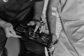
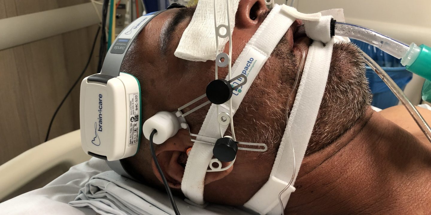
Additionally, the surrogate ICP waveform was recorded using a non-invasive sensor, which allows the monitoring of variations in ICP, in ten-minute sessions that involved the monitoring of other vital signs and manual compression of the internal jugular veins during a minute.
The population was classified into three groups: 21 patients whose skull was kept intact, 21 patients who suffered skull fractures or underwent craniotomy – a surgical procedure that also involves opening the skull to drain hematomas, with immediate closure – and 15 patients who underwent to decompressive craniectomy.
Although all of them showed an increase in ICP during vein compressions, patients in the first two groups also showed an increase in the P2/P1 ratio, used by the research team as an indication of impaired intracranial compliance.
P1 and P2 are two of the three peaks that make up the surrogate ICP waveform. The first peak results from atrial contraction and is followed by a spreading of the volume brought into the cranial mass. Flow transmission through brain tissue produces a second peak that under normal conditions is smoother. In situations where intracranial compliance is impaired, due to the greater resistance offered by internal volumes to blood circulation, the intensity of the first peak is reduced and that of the second increases.
“In this research, we observed that the dynamics of intracranial compliance in patients who underwent craniotomy is similar to that of those who have an intact skull, with a significant elevation of P2. However, in the case of patients who underwent craniectomy surgery, the absence of part of the bone cranial nerve modifies the relationship between the two peaks when we induce an increase in pressure”, ponders Sérgio Brasil.
The article “Intracranial Compliance Assessed by Intracranial Pressure Pulse Waveform” was published on July 2021 in the Brain Sciences journal and is available through the DOI: 10.3390/brainsci11080971
Supporters